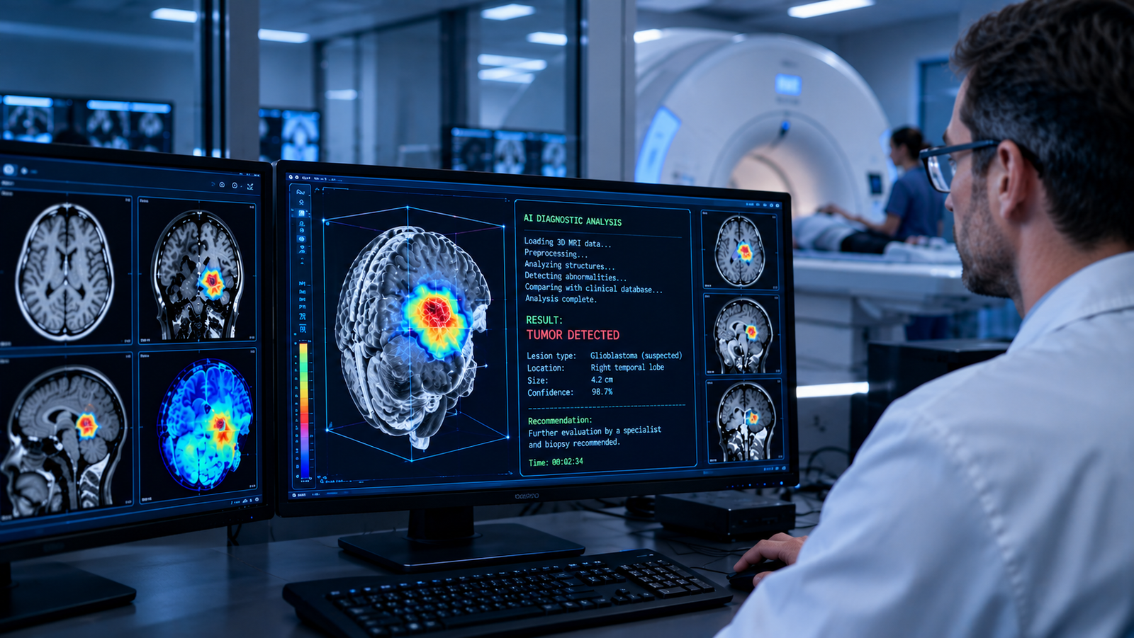

A research team at the Technical University of Munich (TUM) has presented a method capable of partially replacing biopsies with a more modern, albeit currently experimental, approach. At present, according to the developers, the new AI-based program is learning to determine the nature of tumors from CT and MRI scans. In the near future, scientists may be able to include this method in standard cancer diagnostics.
How It Works
The system was named TomoGraphView, and its authors are Professor Jan Peeken and Johannes Kiechle from the Rechts der Isar Hospital. This idea grew out of the quite limited capabilities of diagnostic doctors that all medical radiology faces daily. After all, all the world’s most powerful image recognition models are currently trained on millions of ordinary flat photographs, whereas CT and MRI scans are three-dimensional by nature. Until now, no program had been created that was trained on three-dimensional images, because 3D images take up tens to hundreds of times more server volume and require significant server resources for evaluation.
Therefore, until recently, most scientists managed by splitting three-dimensional images into many two-dimensional ones-from the top, front, and side-and then training existing medical algorithms on these images. The problem is that real tumors are rarely neatly aligned along the cut axes. As soon as a neoplasm is situated at an angle to the usual cut directions, part of the information about its shape, boundaries, and internal structure is simply lost before the computer program even begins to evaluate it.
Researchers Kiechle and Peeken decided to approach computer diagnostics and analysis differently from their predecessors. Instead of three fixed cuts, they are testing a method that evaluates all information around and inside the tumor, analyzing the neoplasm from multiple points evenly distributed across its entire surface. The researchers themselves compare this approach to slicing an apple: if you always cut it in the same direction, part of the internal structures will remain hidden from view, whereas looking from different sides will reveal a much more detailed and reliable picture. TomoGraphView is being tested using the method developed by Kiechle and Peeken, scanning the tumor from all sides to uncover all information about its structure that the human eye simply cannot notice.
Testing the Method on Nearly 2,000 Patients
Before moving to practical application, any new method must always be tested on real patients. The new system was tested on six independent datasets covering scans of nearly 2,000 patients with tumors of the:
- brain;
- breast;
- kidneys;
- liver.
This spread of neoplasm types was not chosen by chance. The testing was meant to show that this analysis is not tuned for just one specific type of tumor, but works as a universal analytical tool. According to the research group, in this comparison with other medical diagnostic programs, TomoGraphView on average demonstrated more accurate results than existing AI systems, and on certain tasks even surpassed the most common trained neural network models specifically created for analyzing 3D medical images.
No Replacement for Biopsy Yet
Despite the compelling nature of the data obtained, the authors of the development are in no rush to make grand statements. It is not a matter of the need for tissue biopsies disappearing in the foreseeable future, since no algorithm can yet compete with histological examination under a microscope. At the Rechts der Isar Hospital, TomoGraphView is viewed differently. The new development is evaluated rather as an additional tool that helps the doctor navigate the volume of data faster and plan further steps more accurately. Talking about completely replacing humans with algorithms is not just premature, but rather foolish. Accelerating the evaluation of received data using this tool will shorten the time to diagnosis, thereby increasing the chances of recovery. Scientists also expect that this method will sharply reduce the number of misdiagnoses, which can not only eliminate lost time or bodily harm from improper treatment, but also reduce treatment costs for the patient while freeing up doctors’ time for other medical tasks.
Where this idea was born is also revealing. From the very beginning, the project was conducted at the intersection of two fields: artificial intelligence research and practical radiation therapy-right where such tools will actually be applied in practice. Developing the tool directly in the clinic is intended by the authors to speed up the technology’s journey from a laboratory prototype to an everyday medical tool.
The project was funded by the Munich-based Wilhelm Sander Foundation, which allocated about 181,000 euros for the study. The results of the work have already been published in the scientific journal Medical Image Analysis.
If the method confirms its reliability in clinical practice, doctors in the future will not have to choose between diagnostic accuracy and unnecessary strain on the patient. A scan that is already performed during a routine examination will be able to provide a detailed diagnosis for which patients previously had to go under the scalpel.